Imaging
����������� Gene expression patterns are documented by taking digital photographs of stained embryos mounted on a slide and examined under microscope. We take two types of digital images.
1. Production snapshots :� Stained embryos in 70% glycerol are transferred from the well in the 96-well plate to a microscope slide. Without applying the coverslip the group of embryos in a drop of glycerol is examined under dissecting microscope (Leica Wild M10 ) and photographed using (ProgRes 3012) digital camera. We usually take two snapshots at two different magnification ( x and x). Production snapshot provide a permanent record of each hybridization event regardless whether or not the experiment was successful. The snapshot images are visible on web reports in cases when no high-resolution images are available.
 �����
����� 
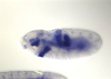
2. High resolution images : Each mounted slide is examined under higher magnification using Zeiss Axiophot and in case an interesting expression pattern is detected we take a large number of high resolution digital photographs using Spot RT digital camera. In most cases individual embryos are photographed using 20X objective so that a single embryo dominates the image field. When necessary we will take a close up image using 40X objective that will highlight fine details of the expression pattern, however in that case the embryo spans beyond the image field. Due to the large-scale nature of our effort we initially did not rigorously orient embryos along the anterior-posterior and dorsal-ventral axis (see FAQ for discussion on image orientation). However we do often rotate the embryo to take a picture of expression pattern from the most favorable angle. Images are stored as JPEGs. On web reports fast-loading low-resolution thumbnails are displayed that can be enlarged to full resolution by clicking.
 ����� �
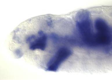
Image staging and grouping
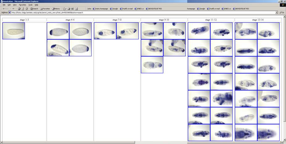
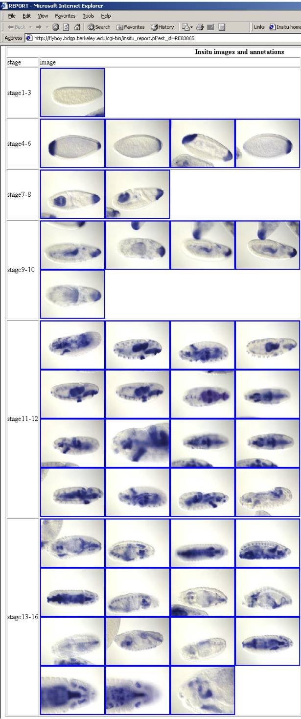
����������� Each captured image is assigned to a specific stage range (see annotation for discussion of stage ranges) and typically multiple images are captured for each stage range. The basic concept is that the group of images assigned to a stage range collectively documents the gene expression pattern at particular stage of development. Individual images usually represent different animals, however in some cases the same animal is photographed several times from different angles or at different focal planes. Highly variable number of images is captured per stage range depending on the complexity of the pattern. Groups of approximately staged images are arranged linearly so that one can follow the development of the pattern across time. During annotation stage-ranges are arranged horizontally so that the pattern unfolds from left to right.
�����������
�����������
����������� On public web reports images are organized vertically and development of the pattern can be followed by scrolling up and down.
����������� �